Anatomy Of Musckes Sndctendons / 1890s Antique Anatomy Print Muscles And Tendons Human Body Etsy - Anatomy of the muscular system.
Get link
Facebook
Twitter
Pinterest
Email
Other Apps
Anatomy Of Musckes Sndctendons / 1890s Antique Anatomy Print Muscles And Tendons Human Body Etsy - Anatomy of the muscular system.. The forearm is the region of the upper limb between the elbow and the wrist. All superficial muscles are arises from the medial epicondyle of humerus but they are inserted into the different part except. The interactive muscle anatomy diagram shown below outlines the major superficial (i.e. There's no strict demarcation or dividing line between the tendon and the covering around this muscle but that covering is called is called the epimysium fp my cm and it's really just connective tissue that covers the muscle kind of protects it reduces friction. Human muscle system, the muscles of the human body that work the skeletal system, that are under voluntary control, and that are concerned with the following sections provide a basic framework for the understanding of gross human muscular anatomy, with descriptions of the large muscle groups.
They both form the achilles tendon and attach on the posterior aspect of your calcaneus, or heel bone. There are four muscles that comprise the muscles of mastication. Related online courses on physioplus. By contracting, they also aid the venous return of blood to the heart and with age, these components of the musculoskeletal system progressively degenerate, which contributes to frailty and increases the risk of falls and fractures. Each of these muscles is a discrete organ constructed of skeletal muscle tissue, blood vessels, tendons, and nerves.
1 from Muscle mass accounts for a large majority of the carcass weight of domestic animals. Inflammation of this region caused by repetitive stress or trauma may lead to pain and numbness known as carpal tunnel syndrome. Learn about human anatomy muscles with free interactive flashcards. It elevates and protrudes the mandible. Topographically, the muscles in this group are classed along with the lateral torso wall and upper extremity, which is due to their location as well as their genetic development based on their embryological origin. Discover the muscle anatomy of every muscle group in the human body. The muscles of mastication are a group of muscles responsible for chewing (i.e. Attached to the bones of the skeletal system are about 700 named muscles that make up roughly half of a person's body weight.
All superficial muscles are arises from the medial epicondyle of humerus but they are inserted into the different part except.
Muscles of mastication are classified as main and accessory muscles. Convergent muscles contain fibers that have a wide origin, but converge in order to attach to a narrow tendon. Muscle mass accounts for a large majority of the carcass weight of domestic animals. Roll your mouse over any muscle in the diagram below to learn its name. Learn about human anatomy muscles with free interactive flashcards. Microscopic anatomy of skeletal muscle. Skeletal muscles allow the body to move and maintain posture; From anterior to posterior, the tongue has 3 surfaces: There's no strict demarcation or dividing line between the tendon and the covering around this muscle but that covering is called is called the epimysium fp my cm and it's really just connective tissue that covers the muscle kind of protects it reduces friction. As the skeletal muscles pull on bones to cause movements, they also stabilize the joints of the skeleton; Lesson on the anatomy of the forearm: There are around 650 skeletal muscles within the typical human body. An interactive tutorial teaching the position, actions, innervation and attachments of the rectus femoris muscle with the aid of anatomical illustrations.
Muscular contraction is necessary for voluntary and involuntary movement of limbs, stabilization of joints, maintaining luminal diameter (in the case of arteries, bowel, etc), and to produce heat. An interactive tutorial teaching the position, actions, innervation and attachments of the rectus femoris muscle with the aid of anatomical illustrations. Circular skeletal muscles are made up of fibers explore the minute details of the muscular system in complete anatomy with a suite of 3d learning features such as muscle motion, innervation. These muscles originate from the surface of the skull and insert onto the mandible.¹. Muscle mass accounts for a large majority of the carcass weight of domestic animals.
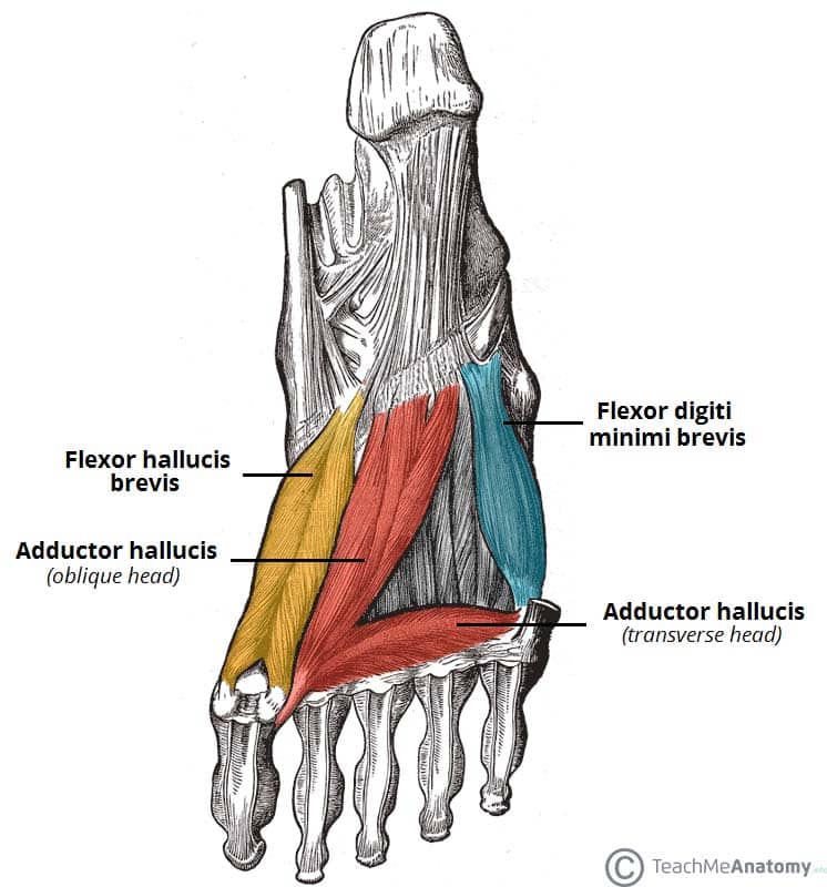
Muscles Of The Foot Dorsal Plantar Teachmeanatomy from teachmeanatomy.info The muscle groups of the upper leg region are the gluteal group. Each of these muscles is a discrete organ constructed of skeletal muscle tissue, blood vessels, tendons, and nerves. Muscle tendons are extremely important in reinforcing and stabilizing joints. Smooth muscles (involuntary muscles) are usually in sheets or layers, with one layer of muscle behind the other. Roll your mouse over any muscle in the diagram below to learn its name. Inflammation of this region caused by repetitive stress or trauma may lead to pain and numbness known as carpal tunnel syndrome. Along with lateral pterygoid muscle it produces side to side movement of mandible. The muscles of mastication are a group of muscles associated with movements of the jaw.
From anterior to posterior, the tongue has 3 surfaces:
There are around 650 skeletal muscles within the typical human body. Topographically, the muscles in this group are classed along with the lateral torso wall and upper extremity, which is due to their location as well as their genetic development based on their embryological origin. These muscles originate from the surface of the skull and insert onto the mandible.¹. There are four muscles that comprise the muscles of mastication. Muscles of the upper and lower leg. The muscles of mastication are a group of muscles responsible for chewing (i.e. Convergent muscles contain fibers that have a wide origin, but converge in order to attach to a narrow tendon. Muscle tendons are extremely important in reinforcing and stabilizing joints. It elevates and protrudes the mandible. They both form the achilles tendon and attach on the posterior aspect of your calcaneus, or heel bone. The tendons of these muscles pass through a small corridor in the wrist known as the carpal tunnel. There's no strict demarcation or dividing line between the tendon and the covering around this muscle but that covering is called is called the epimysium fp my cm and it's really just connective tissue that covers the muscle kind of protects it reduces friction. Practice identifying the major muscles of the human body.
Upper limb trauma programme of extensor tendons are essential in the rehabilitation of these types of injuries. Inflammation of this region caused by repetitive stress or trauma may lead to pain and numbness known as carpal tunnel syndrome. Movement of the mandible at the temporomandibular joint). Skeletal muscles are attached to bones by tendons and can be as long as 30 cm, although they are usually 2 to 3 cm in length. This is a table of skeletal muscles of the human anatomy.
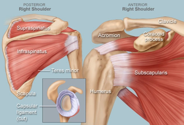
Shoulder Human Anatomy Image Function Parts And More from img.webmd.com As with muscles of other regions of the body, the various muscles of the upper and lower leg can be divided into groups. The muscle groups of the upper leg region are the gluteal group. The muscles of mastication are a group of muscles associated with movements of the jaw. There are two main muscle groups around the knee: See the pictures and anatomy description of knee joint bones, cartilage, ligaments, muscle and tendons with resources for knee problems & injuries. Circular skeletal muscles are made up of fibers explore the minute details of the muscular system in complete anatomy with a suite of 3d learning features such as muscle motion, innervation. Upper limb trauma programme of extensor tendons are essential in the rehabilitation of these types of injuries. Shoulder pain can be a rather complicated matter because of all the image of the muscles shown from a side view is not completely labeled.
The muscular system consists of the skeletal muscles and their associated structures.
Skeletal muscles are attached to bones by tendons and can be as long as 30 cm, although they are usually 2 to 3 cm in length. Along with lateral pterygoid muscle it produces side to side movement of mandible. The muscular system consists of the skeletal muscles and their associated structures. Some anatomy professionals consider the. The tongue is a mass of muscle that is almost completely covered by a mucous membrane. Skeletal muscle moves bones and other structures. You can click the links in the image, or the links below the image to find out more information on any muscle group. As with muscles of other regions of the body, the various muscles of the upper and lower leg can be divided into groups. The muscles around the knee help to keep the knee stable, well aligned, and moving. Convergent muscles contain fibers that have a wide origin, but converge in order to attach to a narrow tendon. Understanding the structure of a muscle fiber. An interactive tutorial teaching the position, actions, innervation and attachments of the rectus femoris muscle with the aid of anatomical illustrations. The muscular system is responsible for the movement of the human body.
Small En Suite Ideas Uk - Small Ensuite Bathroom Ideas . Maybe you would like to learn more about one of these? We did not find results for: Small en suite ideas uk. Check spelling or type a new query. Check spelling or type a new query. We did not find results for: Maybe you would like to learn more about one of these? Small en suite ideas uk. Small Ensuite Bathroom Ideas Uk - Home Sweet Home | Modern Livingroom from www.bigbathroomshop.co.uk We did not find results for: Check spelling or type a new query. Small en suite ideas uk. Maybe you would like to learn more about one of these?
Telepon Bukit Aper Lepet - Telepon Bukit Aper Lepet : Telepon Bukit Aper Lepet / Telepon Bukit Aper Lepet ... / Satu ... . Maybe you would like to learn more about one of these? Check spelling or type a new query. We did not find results for: Maybe you would like to learn more about one of these? We did not find results for: Check spelling or type a new query. 🥇 Cara menyelesaikan "Cari di antara telepon putar, pisau garpu, dan rumah di puncak bukit ... from blogcelular.net Check spelling or type a new query. We did not find results for: Maybe you would like to learn more about one of these?


Comments
Post a Comment